Research and innovation
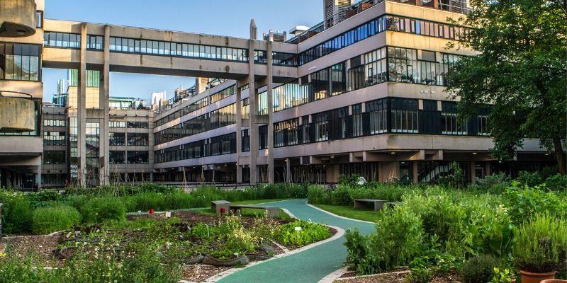
A new home for teaching and research
The Faculty of Biological Sciences is undergoing a major refurbishment to develop its teaching and research capabilities. Creating highly-flexible state of the art laboratories, offices and study space, the investment will provide an exciting new environment for an outstanding student experience and for large multidisciplinary research teams from across multiple faculties to work together.
Research Centres
Below are some of the cross-faculty research centres within the University that draw together expertise, maximising potential for application of novel approaches to solve scientific problems.
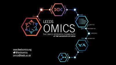
Leeds Omics
Our mission is to engage and unify the critical mass of ‘omics’ researchers at the University of Leeds into one central virtual institute. We have a significant number of active researchers that fit under this “Omics” umbrella.
More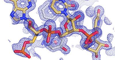
The Astbury Centre
Bringing together researchers from across the University - largely from physics, the biological sciences and chemistry - to allow interdisciplinary approaches to be harnessed to understand the molecular basis of life.
More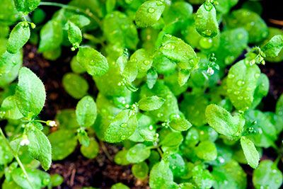
Centre for Plant Sciences
A centre for excellence in plant cell and molecular biology. The Centre has involvement in many European consortia and an extensive range of international collaborations including with the USA, India, Japan and China.
More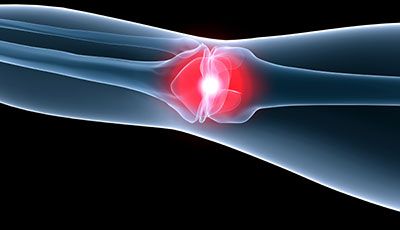
Institute of Medical and Biological Engineering
Focusing on longer lasting joint replacements, tissue sparing interventions and biological scaffolds for tissue regeneration supported by computational and experimental simulation systems for design and pre-clinical testing.
More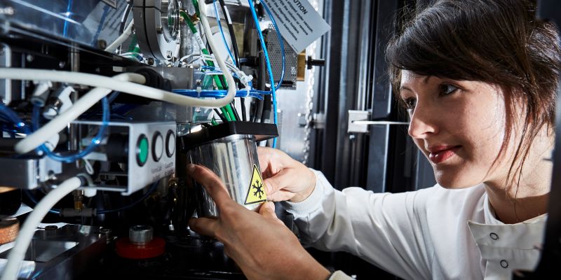
Research Facilities
Our 10 facilities cover the broad research themes of biophysical characterisation, structural elucidation and cellular visualisation. They provide a full pipeline for preparation and complete characterisation of systems from single molecules, to macro-molecular complexes, to cells.
Research Impact
Our research delivers significant benefits to society and the economy. Read through case studies from some of our leading academics that showcase how we have already delivered a high level of impact across the globe as well as exploring our early future impact news stories.
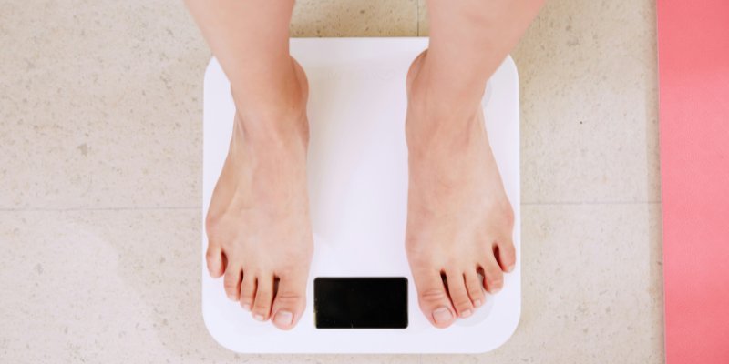
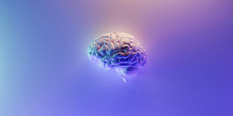
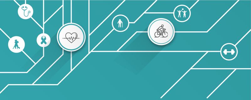
Biomedicine and Health
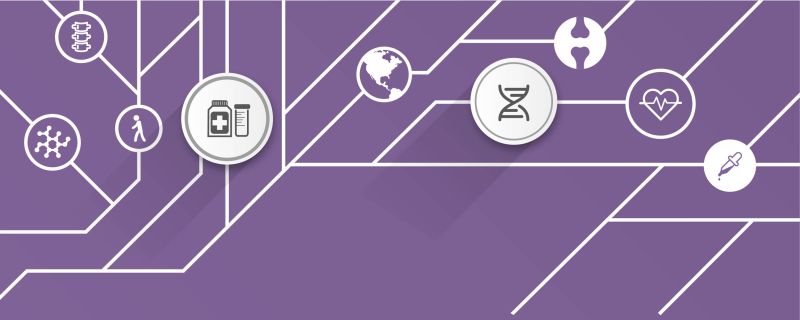
Find out how we are tackling challenges within Biomedicine and Health by reading our impact case studies and some of our other research news.
MoreEcology and Conservation
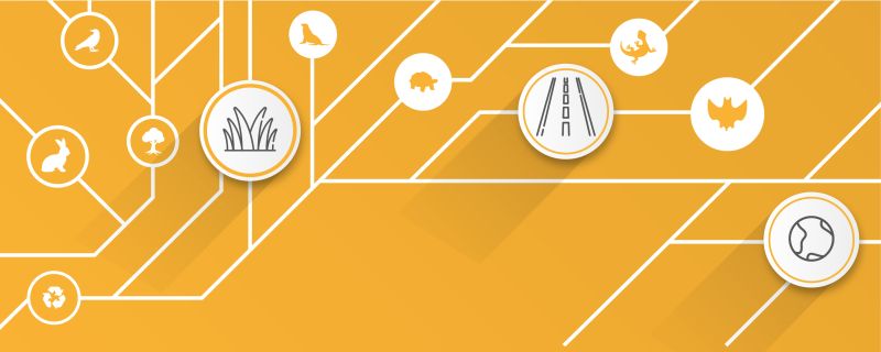
Find out how we are tackling challenges within Ecology and Conservation by reading our impact case studies and some of our other research news.
MoreSport and Exercise Sciences
Find out how we are tackling challenges within Sport and Exercise Science by reading our impact case studies and some of our other research news.
MoreSustainable Agriculture
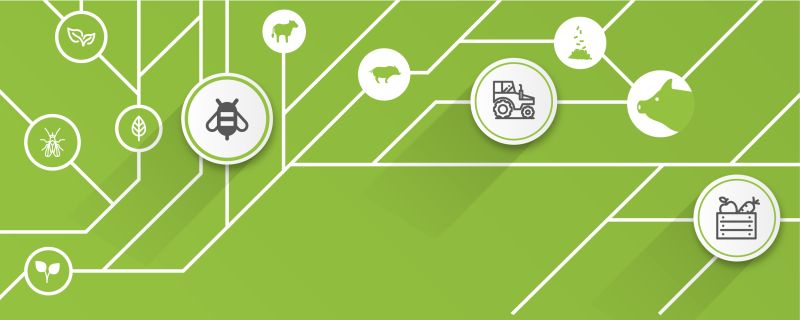
Find out how we are tackling challenges within Sustainable Agriculture by reading our impact case studies and some of our other research news.
More